Anatomy Mapping Foramina : Comprehensive Review Of The Mastoid Foramen Springerlink. Post ethmoidal foramen ant ethmoidal foramen frontoethmoidal suture anterior lacrimal crest optic foramen. Zygote body is a free online 3d anatomy atlas. The video provides a walkthrough of the foramen of the skull (cranial foramina), including the cranial nerves that pass through each foramen. Thoracic nerve roots emerge from the intervertebral foramina into the paravertebral space. In this article we will discuss the anatomy, its contents and clinical relevance of the mandibular mandibular foramen.
In the brain, the interventricular foramina (or foramina of monro) are channels that connect the paired lateral ventricles with the third ventricle at the midline of the brain. Uruj zehra mbbs, mphil, phd last. The floor of the cranial cavity consists of three cranial fossae Prevention of missed anatomy begins with a thorough knowledge of common tooth morphology. Foramen lacerum is an irregular opening located in the middle cranial fossa at the base of the skull.
Duke Neurosciences Lab 2 Spinal Cord Brainstem Surface And Sectional Anatomy from web.duke.edu The structure indicated is the foramen spinosum. The clinician must be aware of the complexities of the root canal system and anatomical variations of the. Prevention of missed anatomy begins with a thorough knowledge of common tooth morphology. The floor of the cranial cavity consists of three cranial fossae Learn vocabulary, terms and more with flashcards, games and other study tools. Thoracic nerve roots emerge from the intervertebral foramina into the paravertebral space. Mobile and tablet users, you can download on appstore or googleplay. It is covered by cartilage after birth.
Thoracic nerve roots emerge from the intervertebral foramina into the paravertebral space.
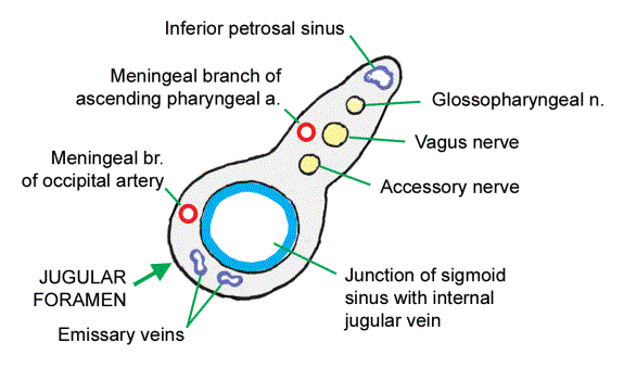
The anatomy of the thoracic outlet extends from the intervertebral foramina and superior. Uruj zehra mbbs, mphil, phd last. See more ideas about anatomy, muscle anatomy, arteries. And sympathetic plexus $u!ular foramen inferior petrosal sinus lossopharyngeal, vagus and. Thoracic nerve roots emerge from the intervertebral foramina into the paravertebral space. The structure indicated is the foramen spinosum. The video provides a walkthrough of the foramen of the skull (cranial foramina), including the cranial nerves that pass through each foramen. The map data is available for public download. Foramen lacerum is an irregular opening located in the middle cranial fossa at the base of the skull. Literature review and pictorial tour. Shahab shahid mbbs • reviewer: The optic foramen, the opening through which the optic nerve runs back into the brain and the large ophthalmic artery enters the orbit, is at the nasal side of the apex. The clinician must be aware of the complexities of the root canal system and anatomical variations of the.
Zygote body is a free online 3d anatomy atlas. Mobile and tablet users, you can download on appstore or googleplay. Foramen lacerum is an irregular opening located in the middle cranial fossa at the base of the skull. Prevention of missed anatomy begins with a thorough knowledge of common tooth morphology. The interventricular foramen, also known as foramen of monro, is part of the ventricular system and the connection between the third ventricle and the lateral ventricle.
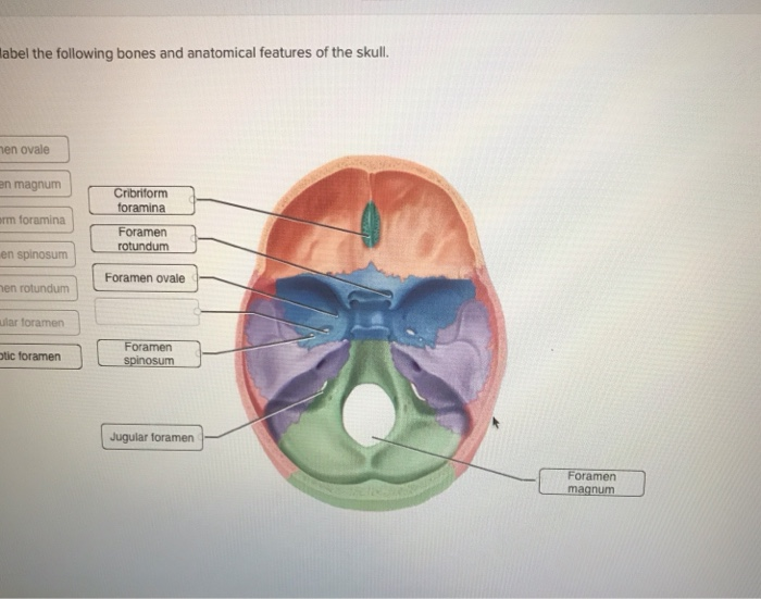
Solved Label The Following Bones And Anatomical Features Chegg Com from media.cheggcdn.com Uruj zehra mbbs, mphil, phd last. Learn vocabulary, terms and more with flashcards, games and other study tools. The map data is available for public download. In the brain, the interventricular foramina (or foramina of monro) are channels that connect the paired lateral ventricles with the third ventricle at the midline of the brain. As channels, they allow cerebrospinal fluid (csf). Thoracic nerve roots emerge from the intervertebral foramina into the paravertebral space. Mobile and tablet users, you can download on appstore or googleplay. Zygote body is a free online 3d anatomy atlas.
The floor of the cranial cavity consists of three cranial fossae
The structure indicated is the foramen spinosum. Uruj zehra mbbs, mphil, phd last. In this article we will discuss the anatomy, its contents and clinical relevance of the mandibular mandibular foramen. Post ethmoidal foramen ant ethmoidal foramen frontoethmoidal suture anterior lacrimal crest optic foramen. The clinician must be aware of the complexities of the root canal system and anatomical variations of the. This is the revised version of the there are twelve cranial nerves, which leave the brain and pass through foramina in the skull. The journal of laryngology clinical anatomy of blockade of the pterygopalatine ganglion: Information on the foramen spinosum by the anatomyzone daily feed. Zygote body is a free online 3d anatomy atlas. This article discusses each of the aforementioned fossae and their associated foramina. Literature review and pictorial tour. The map data is available for public download. The floor of the cranial cavity consists of three cranial fossae
Thoracic nerve roots emerge from the intervertebral foramina into the paravertebral space. This article discusses each of the aforementioned fossae and their associated foramina. View, isolate, and learn human anatomy structures with zygote body. As channels, they allow cerebrospinal fluid (csf). Shahab shahid mbbs • reviewer:
Structures Passing Through Jugular Foramen Mnemonic Epomedicine from epomedicine.com The optic foramen, the opening through which the optic nerve runs back into the brain and the large ophthalmic artery enters the orbit, is at the nasal side of the apex. It is formed by the apex of the petrous temporal bone and allows the. This is the revised version of the there are twelve cranial nerves, which leave the brain and pass through foramina in the skull. The structure indicated is the foramen spinosum. Literature review and pictorial tour. Mobile and tablet users, you can download on appstore or googleplay. Zygote body is a free online 3d anatomy atlas. The video provides a walkthrough of the foramen of the skull (cranial foramina), including the cranial nerves that pass through each foramen.
The interventricular foramen, also known as foramen of monro, is part of the ventricular system and the connection between the third ventricle and the lateral ventricle.
Literature review and pictorial tour. The anatomy of the thoracic outlet extends from the intervertebral foramina and superior. Lingual foramina, anatomy, mandibular midline | the purpose of the present study was to determine the incidence, size, location, course. View, isolate, and learn human anatomy structures with zygote body. Foramen lacerum is an irregular opening located in the middle cranial fossa at the base of the skull. The optic foramen, the opening through which the optic nerve runs back into the brain and the large ophthalmic artery enters the orbit, is at the nasal side of the apex. Prevention of missed anatomy begins with a thorough knowledge of common tooth morphology. It is covered by cartilage after birth. The video provides a walkthrough of the foramen of the skull (cranial foramina), including the cranial nerves that pass through each foramen. Each fossa contains specific foramina, through which various anatomical structures pass through. The clinician must be aware of the complexities of the root canal system and anatomical variations of the. See more ideas about anatomy, muscle anatomy, arteries. It is formed by the apex of the petrous temporal bone and allows the.
The map data is available for public download anatomy map. Origins, pathways & basic applied anatomy.
Share :
Post a Comment
for "Anatomy Mapping Foramina : Comprehensive Review Of The Mastoid Foramen Springerlink"


Post a Comment for "Anatomy Mapping Foramina : Comprehensive Review Of The Mastoid Foramen Springerlink"